At first I was quite depressed to hear this. They provided me with the MRI pictures on cd so I began to view them myself on my computer.
What I found however, was not in line with what they were saying. To go from 16mm to 27mm would have been almost double in size.
I measured the distance between the edge of my breast to the nipple and it is about 3 inches or 76.2 mm. A lump that is 27mm is about 1/3 of the distance that is between nipple and chest on my breast. When I look at the images I see the reverse situation - that the lump appears to be larger on the July MRI and closer to 1/3 of the "real estate" between beginning of breast curve and nipple.
I am inserting the MRI pics of note here.
The first one is the first MRI of the lump from July 5th of 2006:
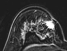
The next several are from the most recent MRI on August 25th - 6 weeks after the first MRI:


What I notice here are several things. The contrast is lighter on the second MRI for the lump. Is that because of a different contrast medium OR is it because there is less of a blood supply in the tumor? Hmmm?
And I notice it seems to be retreating from the surface of the skin which is why the skin retraction is softening. I sure wish I could find out more about skin retraction - why it happens and why it stops happening. But to me, the second MRI is actually encouraging.

No comments:
Post a Comment